Image To
Intervention
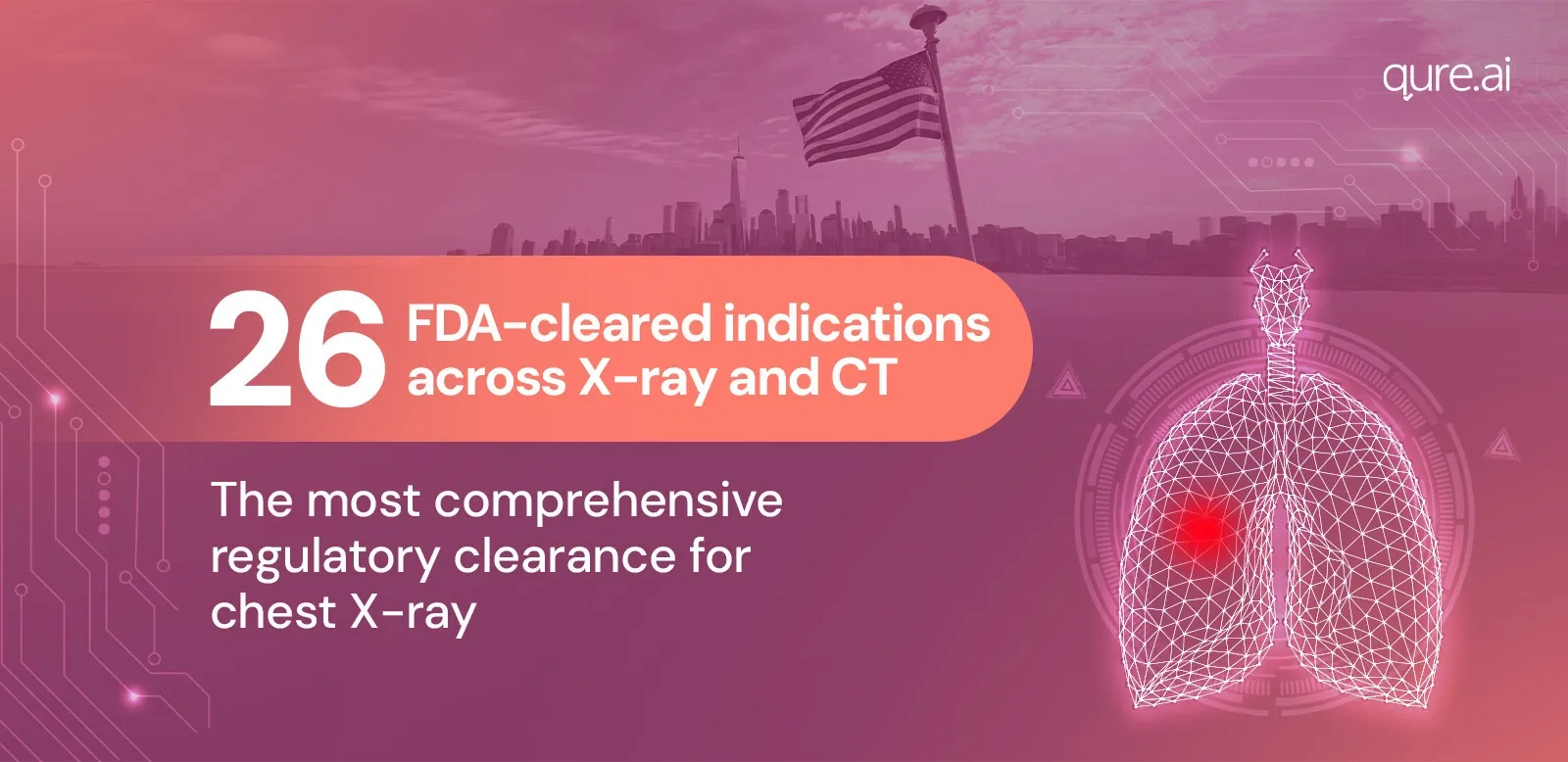
End-to-end healthcare AI for early lung nodule care
Qure.ai supports Radiologists, Pulmonologists and Emergency Physicians across the lung nodule pathway - Identify at-risk patients based on imaging, enabling faster access to treatment across real-world clinical workflows


For conditions such as intracranial hemorrhage, time is of the essence and those precious minutes can be life-changing for our patients. We have done extensive validation of the Qure.ai qER solution and are excited to continue to partner with Qure.ai and improve care for our patients.
Benjamin W. Strong
MD and Chief Medical Officer
vRad

A multicenter publication on missed and mislabeled chest radiography findings including pneumothoraces and pleural effusions reported up to 96% sensitivity and 100% specificity for the qXR algorithm.
Dr. Subba Digumarthy, MD
Attending Radiologist, Thoracic Imaging, Massachusetts General Hospital
Associate Professor, Harvard Medical School

Medical imaging AI holds immense potential in the battle against lung cancer in the United States. It is great to see the breadth of FDA clearances rolling in to enable the exploration and activation of algorithms that can support radiologists and pulmonologists. This will help to detect lung nodules earlier using chest X-ray, and also analyze them in detail on chest CT.
Dr. Javier Zulueta
Professor of Medicine, former Chief of Pulmonary, Critical Care, and Sleep Division at Mount Sinai Morningside Hospital.

AI serves as an additional set of eyes for radiologists, enhancing detection by flagging lung nodules that may require further evaluation. This AI-driven approach may aid in identifying more nodules which we hope supports patient care and enables us to evaluate the broader impact of medical imaging AI. The clinical trial will evaluate how many patients require follow-up CT scans, biopsies, and how many more lung cancer cases are diagnosed earlier using AI. The hope is that this clinical trial will not only advance early detection but also drive meaningful transformation in lung cancer surveillance
Dr. Amit Gupta
Cardiothoracic Radiologist and Modality Director of Diagnostic Radiography at University Hospitals Cleveland Medical Center

I’ve had the opportunity to work extensively with qXR and qCT in real lung nodule workflows through our Sinai Chicago collaboration with Qure.ai. qXR’s ability to detect subtle nodules without creating reader fatigue has been particularly impactful. And on the qCT side, collaborating directly with Qure’s product engineers to refine the annotation tools has resulted in a workflow that truly supports both radiologists and referring physicians. It’s rewarding to contribute to a solution designed with the end user and our patients in mind.
Dr Amar P. Shah
MD - System Chair of Radiology, Sinai Chicago, Chicago IL

For conditions such as intracranial hemorrhage, time is of the essence and those precious minutes can be life-changing for our patients. We have done extensive validation of the Qure.ai qER solution and are excited to continue to partner with Qure.ai and improve care for our patients.
Benjamin W. Strong
MD and Chief Medical Officer
vRad
A multicenter publication on missed and mislabeled chest radiography findings including pneumothoraces and pleural effusions reported up to 96% sensitivity and 100% specificity for the qXR algorithm.
Dr. Subba Digumarthy, MD
Attending Radiologist, Thoracic Imaging, Massachusetts General Hospital
Associate Professor, Harvard Medical School
Medical imaging AI holds immense potential in the battle against lung cancer in the United States. It is great to see the breadth of FDA clearances rolling in to enable the exploration and activation of algorithms that can support radiologists and pulmonologists. This will help to detect lung nodules earlier using chest X-ray, and also analyze them in detail on chest CT.
Dr. Javier Zulueta
Professor of Medicine, former Chief of Pulmonary, Critical Care, and Sleep Division at Mount Sinai Morningside Hospital.
AI serves as an additional set of eyes for radiologists, enhancing detection by flagging lung nodules that may require further evaluation. This AI-driven approach may aid in identifying more nodules which we hope supports patient care and enables us to evaluate the broader impact of medical imaging AI. The clinical trial will evaluate how many patients require follow-up CT scans, biopsies, and how many more lung cancer cases are diagnosed earlier using AI. The hope is that this clinical trial will not only advance early detection but also drive meaningful transformation in lung cancer surveillance
Dr. Amit Gupta
Cardiothoracic Radiologist and Modality Director of Diagnostic Radiography at University Hospitals Cleveland Medical Center
I’ve had the opportunity to work extensively with qXR and qCT in real lung nodule workflows through our Sinai Chicago collaboration with Qure.ai. qXR’s ability to detect subtle nodules without creating reader fatigue has been particularly impactful. And on the qCT side, collaborating directly with Qure’s product engineers to refine the annotation tools has resulted in a workflow that truly supports both radiologists and referring physicians. It’s rewarding to contribute to a solution designed with the end user and our patients in mind.
Dr Amar P. Shah
MD - System Chair of Radiology, Sinai Chicago, Chicago IL
AI solutions in healthcare trusted globally


The only platform unifying detection and follow-up
Build detection and navigation in a single workflow. Start where you are, upgrade anytime.

Imaging & Interpretation
Seamlessly integrates with PACS, EMR, dictation, and local reporting systems. Built to accelerate interpretation without changing how you work
qXR - LN : Identify lung nodules on Chest Radiographs
Detect and localise nodules on chest radiographs
- Find subtle pulmonary nodules (6-10mm) with high accuracy
- Expand IPN programs - Unlock additional revenue
- 1 lung nodule found in 1120 CXRs as per on-ground evidence*
Chest radiograph lung nodule detection & localization (qXR-LN) FDA Cleared for Radiologists, pulmonologists, and emergency physicians reviewing adult chest - Learn more →
*Union 2024 Vietnam Abstract
qCT LN Quant : Automate nodule Quantification on CTs
Lung Nodule Quantification & Volumetry on CTs
- Precisely quantify nodules with Volumetric Segmentation
- Avail reimbursement under 0722T/0721T codes
- Access Lung‑RADS® & Brock score based risk stratification* and follow-up recommendation based on Fleischner guidelines
Advanced CT-based nodule quantification and tracking (qCT LN Quant) FDA Cleared for radiologists and pulmonologists - Learn more →
*Risk stratification by the intended users

Follow-up Management Platform - qTrack
Impact every patient touchpoint with qTrack. Less administrative burden. More time for patient care
Structured case finding and longitudinal tracking
- Supports radiology workflow orchestration
- Automates longitudinal tracking across imaging and follow-up
- Reduce FTE hours through AI-assisted workflows
Giving time back to radiologists
- Track patient follow-up status in real-time
- Avoid duplicate or manual data entry
- Communicate timely with AI-powered care coordination
How LLMs Go Beyond Traditional NLP

NLP
Rule-first Processing
Treats all nodules the same without anatomy, risk, or context.

Relies on predefined rules & keywords lists

Scans reports for exact matches, ignores critical context

Leads to poor outcomes

LLM
Reason-first Intelligence
Extracts context to assess true risk basis EMRs.

Integrates multiple data sources

Understands full clinical context

Handles synonyms, nuance, report styles

Reimbursement
Reimbursement for AI-enabled lung cancer and neurocritical care pathways is evolving. Qure’s products within the lung cancer and neuro pathways are billable under CMS-issued CPT codes (such as 0721T, 0727T), allowing hospitals to establish a stable revenue model while optimally utilizing healthcare AI solutions to improve accurate and faster diagnostics. Qure works with revenue-cycle teams to align documentation, coding, and payer engagement within existing imaging and follow-up workflows.
Please consult local medical-necessity and documentation policies before billing and prior authorization.
For more information please write to us at partner@qure.ai
Saving Lives with AI: How Early Lung Cancer Detection Changed Diane’s Life
Click to Watch the Story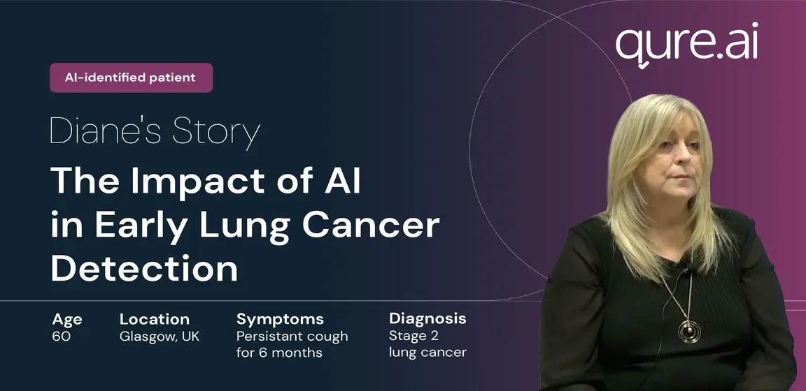
News