South Africa has a high mortality rate for Tuberculosis as well as Lung cancer due to late detection and lack of screening programs for these diseases.

Back
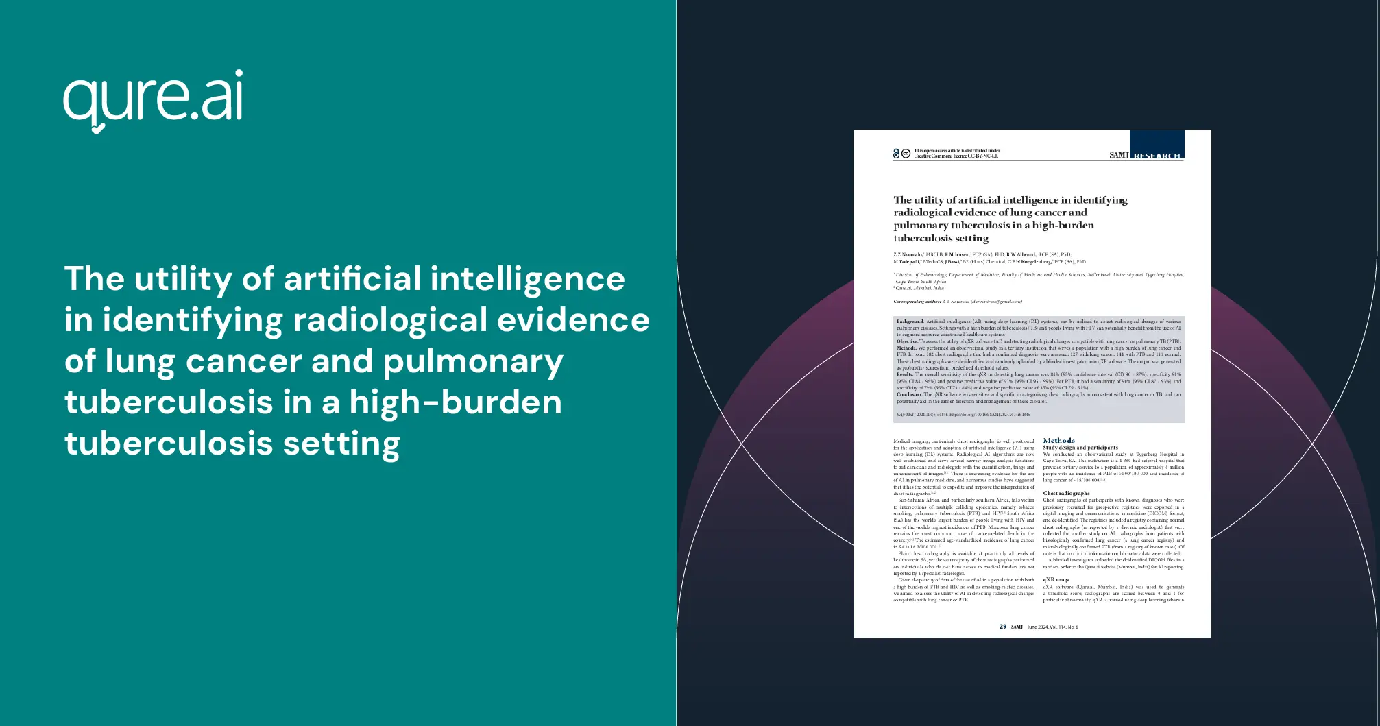
Our latest study published in the South Africa Medical Journal titled "The Utility of Artificial Intelligence in Identifying Radiological Evidence of Lung Cancer and Pulmonary Tuberculosis in a high-burden Tuberculosis Setting"in collaboration with Stellenbosch University and Tygerberg Hospital, South Africa states how Qure's AI Solution, qXR - for lung health, can help detect multiple pulmonary diseases with the same infrastructure available in the local hospitals in South Africa.
The AI solutions integrates seamlessly into old hardware (retrofit) models, making it easier for healthcare centres with low resources to avail the best in class technology. Qure is working towards health system strengthening across the globe and ensuring health equity to the last mile.
Background
Artificial intelligence (AI), using deep learning (DL) systems, can be utilised to detect radiological changes of various pulmonary diseases. Settings with a high burden of tuberculosis (TB) and people living with HIV can potentially benefit from the use of AI to augment resource-constrained healthcare systems.
Objective
To assess the utility of qXR software (AI) in detecting radiological changes compatible with lung cancer or pulmonary TB (PTB).
Methods
We performed an observational study in a tertiary institution that serves a population with a high burden of lung cancer and PTB. In total, 382 chest radiographs that had a confirmed diagnosis were assessed: 127 with lung cancer, 144 with PTB and 111 normal. These chest radiographs were de-identified and randomly uploaded by a blinded investigator into qXR software. The output was generated as probability scores from predefined threshold values.
Results
The overall sensitivity of the qXR in detecting lung cancer was 84% (95% confidence interval (CI) 80 - 87%), specificity 91% (95% CI 84 - 96%) and positive predictive value of 97% (95% CI 95 - 99%). For PTB, it had a sensitivity of 90% (95% CI 87 - 93%) and specificity of 79% (95% CI 73 - 84%) and negative predictive value of 85% (95% CI 79 - 91%).
Conclusion
The qXR software was sensitive and specific in categorising chest radiographs as consistent with lung cancer or TB, and can potentially aid in the earlier detection and management of these diseases.