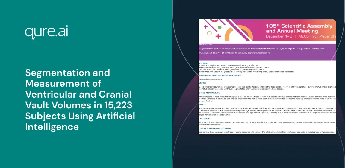
PURPOSE
Back
True volumetric measurement of the cerebral ventricles could potentially improve the diagnosis and follow-up of hydrocephalus. However, manual image segmentation is an impractically laborious process to be applied routinely. In this work, we utilize deep learning algorithms to provide an automated solution for precise ventricular segmentation and volume quantification in a large dataset.
METHOD AND MATERIALS
A large database of head computed tomographic (CT) scans was utilized to train and validate convoluted neural network models. Lateral ventricles were manually annotated in 103 scans and were randomly split with a training-validation ratio of 4:1. One U-net model was trained to segment the lateral ventricles in each slice, and another to segment the cranial vault. Each model was validated against the manually annotated images using the DICE index. Both networks were then used to segment and quantify the ventricular and cranial vault volumes of a large random sample from our database.
RESULTS
Both the ventricular volume and the cranial vault U-net models showed high fidelity to the manual annotation (DICE 0.909 and 0.983, respectively). They were then applied to a subset of 15,223 head CT scans from our database. Among these scans, 1,999 (13.1%) had a radiological report of cerebral atrophy and 1,404 (9.2%) of hydrocephalus. Age median was 40 years and 41.5% were females. Patients reported to have cerebral atrophy were confirmed by the U-nets to have larger ventricles. Cranial vault volume increased until the age group of 10-20, after which a plateau was observed. Conversely, ventricular volume increased with age without a plateau, evidently due to cerebral atrophy. Males had 13% larger cranial vault volumes than females. The ventricle-to-vault ratio showed no differences between the two sexes in younger ages, but men had a steeper increase with age than women.
CONCLUSION
This is the first study to measure ventricular volumes in such a large dataset, which has been made possible using artificial intelligence. Here we provide a robust method to establish normal values for ventricular volumes and a tool to routinely report these volumes on CT scans and evaluate for hydrocephalus.
CLINICAL RELEVANCE/APPLICATION
Deep learning tools can provide ventricular volume measurements on head CTs efficiently and with high fidelity, and can assist in the diagnosis of hydrocephalus.