Background
Published 05 Jan 2021
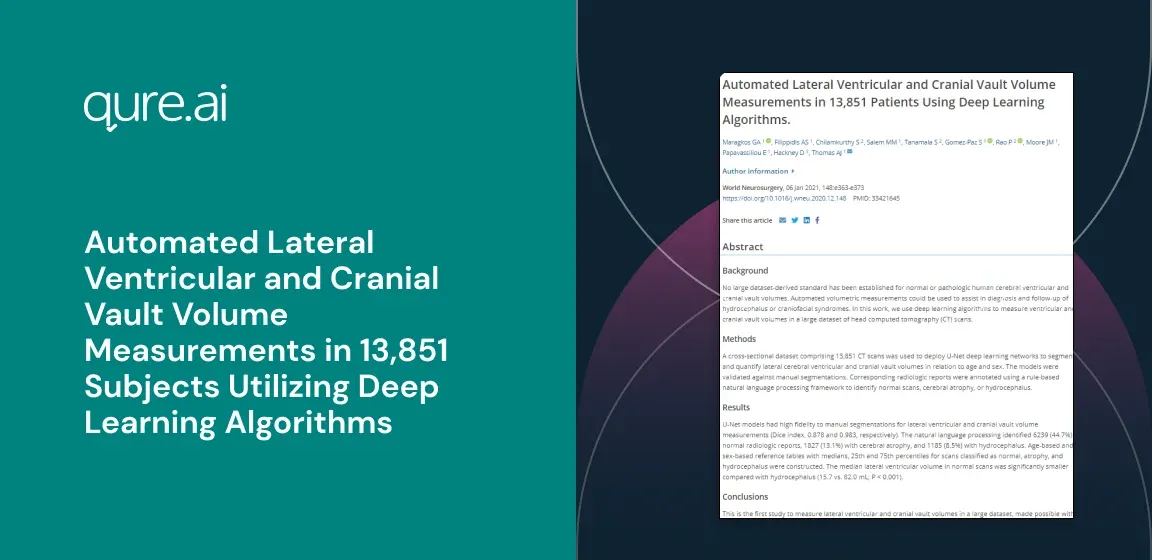
Automated Lateral Ventricular and Cranial Vault Volume Measurements in 13,851 Subjects Utilizing Deep Learning Algorithms
Author: Georgios A Maragkos 1, Aristotelis S Filippidis 1, Sasank Chilamkurthy 2, Mohamed M Salem 1, Swetha Tanamala 2, Santiago Gomez-Paz 1, Pooja Rao 2, Justin M Moore 1, Efstathios Papavassiliou 1, David Hackney 3, Ajith J Thomas 11
Back
Currently, no large dataset-derived standard has been established for normal or pathologic human cerebral ventricular and cranial vault volumes. Automated volumetric measurements could be used to assist in diagnosis and follow-up of hydrocephalus or craniofacial syndromes. In this work we use deep learning algorithms to measure ventricular and cranial vault volumes in a large dataset of head computed tomography (CT) scans.
Methods
A cross-sectional dataset comprising 13,851 CT scans was utilized to deploy U-net deep learning networks to segment and quantify lateral cerebral ventricular and cranial vault volumes in relation to age and sex. The models were validated against manual segmentations. Corresponding radiological reports were annotated using a rule-based natural language processing (NLP) framework to identify normal scans, cerebral atrophy, or hydrocephalus.
Results
U-net models had high fidelity to manual segmentations for lateral ventricular and cranial vault volume measurements (DICE 0.878 and 0.983, respectively). The NLP identified 6,239 (44.7%) normal radiological reports, 1,827 (13.1%) with cerebral atrophy and 1,185 (8.5%) with hydrocephalus. Age- and sex-based reference tables with medians, 25th and 75th percentiles for scans classified as normal, atrophy and hydrocephalus were constructed. The median lateral ventricular volume in normal scans was significantly smaller compared to hydrocephalus (15.7mL vs 82.0mL, P<0.001).
Conclusion
This is the first study to measure lateral ventricular and cranial vault volumes in a large dataset, made possible with artificial intelligence. We provide a robust method to establish normal values for these volumes and a tool to report these on CT scans when evaluating for hydrocephalus.
Authors
Georgios A Maragkos 1, Aristotelis S Filippidis 1, Sasank Chilamkurthy 2, Mohamed M Salem 1, Swetha Tanamala 2, Santiago Gomez-Paz 1, Pooja Rao 2, Justin M Moore 1, Efstathios Papavassiliou 1, David Hackney 3, Ajith J Thomas 11
Citation
1. Neurosurgery Service 2. Beth Israel Deaconess Medical Center 3. Harvard Medical School 4. Boston 5. MA 6. USA 7. Qure.ai 8. Mumbai 9. India 10. Radiology Department 11. Beth Israel Deaconess Medical Center 12. Harvard Medical School 13. Boston 14. MA 15. USA