The Benefits of AI in Neuro-Critical Radiology
Back
AI's integration into radiology is notably substantial, with 75% of over 500 FDA-cleared AI algorithms targeting radiology practice, an increase from 70% in 2021. This increasing presence of AI in radiology indicates its potential to revolutionize not just radiology in general but also neuroradiology.
One of the core benefits of AI in neuroradiology is its promise to enhance efficiency and reduce errors. By improving data processing and image interpretation, AI aids radiologists in elevating the quality of patient care.
These improvements are not just limited to diagnostics but also various neuroimaging aspects. Current AI applications in neuroradiology include worklist prioritization, lesion detection, anatomical segmentation, safety, quality, and precision education, encompassing a range of diseases from neurodegeneration to trauma.
The potential of AI in neuroradiology is vast, contributing significantly to the accuracy and efficiency in many facets of this field. It offers substantial opportunities for insights into brain pathophysiology, developing models for treatment decisions, and enhancing current prognostic and diagnostic algorithms.
Six clinical objectives supported by AI in radiology have been identified: more efficient workflow, shortened reading time, reduced dosage of contrast agents, earlier disease detection, improved diagnostic accuracy, and more personalized diagnostics. These objectives encapsulate the broad spectrum of benefits that AI brings to neuroradiology.
The exponential growth in AI applications in medical imaging, particularly in neuroradiology, is noteworthy. From about 200 peer-reviewed publications in 2010 to approximately 1000 in 2019, neuroradiology accounts for one-third of these articles. This reflects the field's suitability for AI applications due to its rich, multidimensional, and multimodal data.
Various AI-enabled clinical applications have been introduced in neuroradiology, spanning disease detection, lesion quantification, segmentation, image reconstruction, outcome prediction, workflow optimization, and scientific discovery.
These applications are poised to become mainstream in routine clinical practice, with workflow optimization tools likely to precede deep learning–based computer-aided diagnosis tools.
AI's role in detecting irregularities on imaging, particularly in identifying urgent findings like intracranial hemorrhage, acute infarction, large-vessel occlusion, aneurysm detection, and traumatic brain injury, is crucial.
These AI applications assist in worklist prioritization and disease quantification, aiding in tasks like 3D reconstructions, segmentation, and identifying new lesions.
Finally, deep learning proves remarkably effective in reconstructing diagnostic-quality images and removing artifacts, even under extreme protocol parameters.
This is significant in rapidly accelerated MR imaging, ultralow radiation dose CT, and nuclear medicine acquisitions. Such advancements will change the potential indications for lengthy and radiation-heavy studies, opening doors to high-fidelity dynamic imaging.
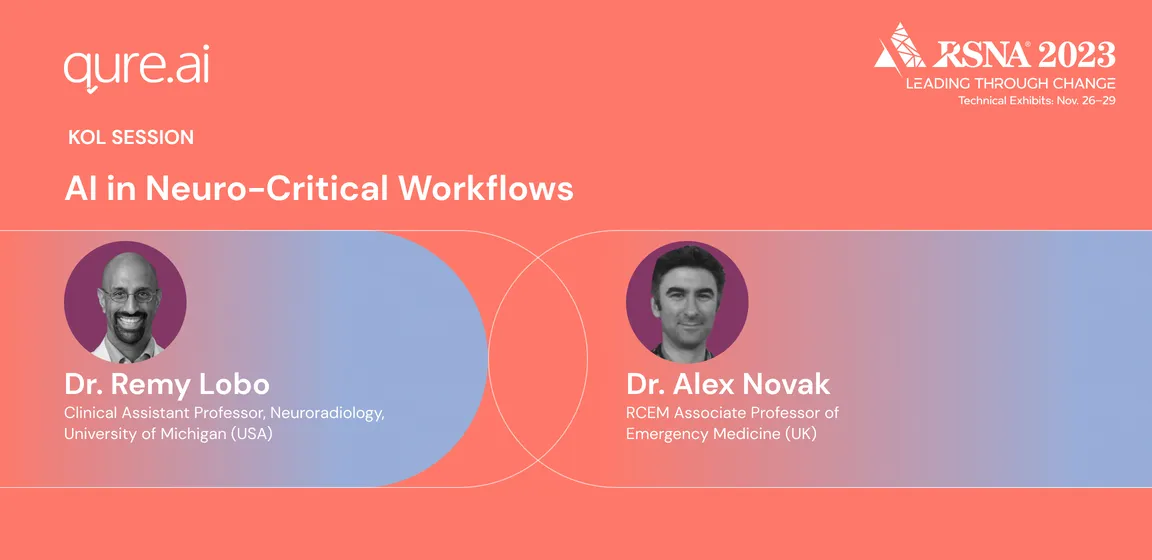
This brings us to an exciting event at the RSNA 2023 that promises to delve deeper into this topic: Booth Session 4, titled "AI in Neuro-Critical Workflows."
Scheduled for Nov 29, 2023, Wednesday at 11:00 am CST, this session is a must-attend for anyone interested in the intersection of AI and neuro-critical care.
Dr. Remy Lobo, a Clinical Assistant Professor of Neuroradiology at the University of Michigan (USA), and Dr. Alex Novak, RCEM Associate Professor of Emergency Medicine (UK), will lead this insightful discussion. They will explore the latest advancements in AI that are reshaping critical neurological care, drawing on their extensive experience and research in the field.
