Shamie Kumar describes her perspective on how radiography has evolved over time, the impact radiographers can have in detecting abnormal X-rays and reflects how she views fast approaching AI in advancing current skills.

Back
The red dot system
Often one of the first courses a newly qualified radiographer attends is the red dot course. This course demonstrates pathologies and abnormalities often seen in x-rays some obvious, others not, giving radiographers the confidence to alert the referring clinician and/or radiologist that there is something abnormal they have seen.
The red dot system is a human alert system, often 2 pairs of eyes are better than one and assist with near misses. How this is done in practice can vary between hospitals, in the era of films the radiographer would place a red dot sticker on the film itself before returning it to clinician or radiologist. In the world of digital imaging this is often done during ‘post documentation’ a term used once the x-ray is finished, the radiographer will complete the rest of the patient documentation to suggest the x-ray is complete, ready to be viewed and reported. As part of this process the radiographer can change the status of the patient to urgent along with a note for what has been observed. From this the radiologist knows the radiographer has seen something urgent on the image and the patient appears at the top of their worklist for reporting and, so the radiologist can view the radiographer’s notes.
The Role of AI in Radiology
Artificial Intelligence (AI) is moving at a pace within healthcare and fast approaching radiology departments, with algorithms showing significant image recognition in detecting, characterisation and monitoring of various diseases within radiology. AI excels in automatically recognising complex patterns in imaging data providing quantitative assessments of radiological characteristics. With the numbers for diagnostic imaging requests forever increasing, many AI companies are focusing on how to ease this burden and supporting healthcare professionals.
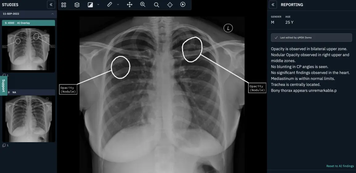
AI triage is done by the algorithm based on abnormal and normal finding’s this is used to create an alert for the referring clinician/radiologist. It can be customised to the radiologist, for example colour-coded flags, red for abnormal, green for normal, patients with a red flag would appear at the top of the radiologist worklist. For the referring clinicians who don’t have access to the reporting worklist, the triage would be viewed on the image itself with an additional text note suggesting abnormal or normal.
What does AI do that a radiographer doesn’t already? AI is structured in the way it gives the findings for example a pre-populated report with its findings or an impression summary and its consistent without reader variability. So, the question now becomes what AI can do beyond the red dot system, here the explanation is straightforward, often a radiographer wouldn’t go to the extent of trying to name what they have seen, especially in more complex x-rays like the chest where there are multiple structures and pathologies. For example a radiographer would mention, right lower lobe and may not go beyond this, often due to confidence and level of experience.
AI can fill this gap, it can empower radiographers and other healthcare professionals with its classification of pathologies identifying exactly what has been identified on the image, based on research and training of billions of data sets with high accuracy.
The radiographers may have the upper hand with reading the clinical indication on the request form and seeing the patient physically, which undoubtable is of significant value, however the red dot system has many variables specific to that individual radiographer’s skills and understanding. It is also limited to giving details of what they have noted to just the radiologist, what about the referring clinician who doesn’t have access to the radiology information system (RIS) where the alert and notes are? Do some radiographers add a text note on the x-ray itself?
Summary
Yes, AI is a technological advancement of the red dot system and will continue to evolve. It is structured in how it gives the findings, it does this consistently with confidence. Adding value to early intervention, accurate patient diagnosis, contributing to reducing misdiagnosis and near misses. AI is empowering radiographers, radiologist, referring clinicians and junior doctors by enhancing and leveraging their current knowledge to a level where there is consistent alerts and classified findings that can even be learned from. This doesn’t replace the red dot system but indeed enhances it.
The unique value a radiographer adds to the patient care, experience and physical interaction can easily be supplemented with AI, allowing them to alert with confidence and manage patients, focusing the clinician time more effectively.
About Shamie Kumar
Shamie Kumar is a practicing HCPC Diagnostic Radiographer; graduating from City University London, BSc Honors in Diagnostic Radiography in 2009 and part of Society of Radiographers with over 10 years of clinical knowledge and skills within all aspects of radiography. She studied further in leadership, management and counselling with a keen interest in artificial intelligence in radiology.